Computer Diagnostics
- Vision check-up
- Examinations
- Investigations
- Tests
- Contact Lenses
- Eyeglasses fitting
--------
Vision Check-Up (Pretest of eyesight)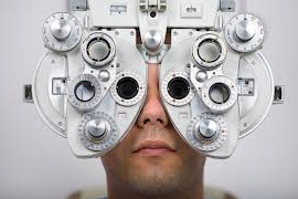
Our experienced optometrists make patient’s eyesight check up. It is not just helping a patient to know the eyesight condition, but sometimes it could also help to identify other health problems, to detect other health-related conditions such as diabetes and high blood pressure. An eye tests can ensure your prescription is up-to-date, and monitor early signs of eye disease, including glaucoma and cataract. For most people this is every two years to make an eyesight check up. Usually it includes:
- Visometry (Checking of visual acuity with correction and without it);
- Determination of refraction's condition;
- Measurement of intraocular pressure /IOP/ (if necessary);
- Determination of parameters of eye glasses or contacts.
Examinations
Pupil function. An examination of pupillary function includes inspecting the pupils for equal size (1 mm or less of difference may be normal), regular shape, reactivity to light, and direct and consensual accommodation.
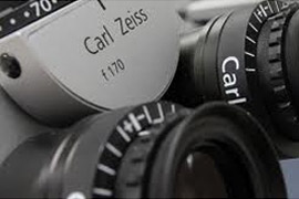
Biomicroscopy with a slit lamp with large magnification the doctor examines the structure of a frontal part of the eyes - eyelids, fibers, the mucous, the iris, the crystalline lens and the cornea.
Ophthalmoscopy - with the help of bright concentrated ray of light under large magnification the doctor examines the internal part of an eyeball. Ophtalmoscopy is one of the basic methods used in the examination of the retina
Perimetry(Visual fields testing, Automated perimetry exam). Perimetry or visual field test is an eye examination that can detect dysfunction in central and peripheral vision. These changes could be caused by various medical conditions such as glaucoma, stroke, brain tumours or other neurological deficits. There are several methods of this examination. It could be performed by an eye specialist using equipment or completely automated. Perimetry is the systematic measurement of visual field function. There are two most commonly used types of perimetry, they are kinetic perimetry and threshold static automated perimetry. Evaluation of central visual field plays the significant role in diagnostic and dynamic observation of patients with glaucoma. Computer statistic perimetry has recently become the most popular method of visual fields estimation.
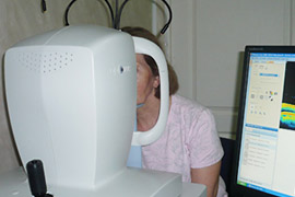
Tonometry (Intraocular Pressure (IOP) measurement), this is the procedure which eye doctors perform to determine the intraocular pressure. It is one of the very important tests in the evaluation of patient’s eyesight condition. There are several methods of tonometry, they could be contact and non-contact. In our clinic we could offer different techniques of this examination, but the most popular between patients is non-contact pneumotonometry.
A pneumotonometer utilizes a pneumatic sensor. Filtered air is pumped into the piston and travels through a small (5-mm.) fenestrated membrane at one end. This membrane is placed against the cornea. The balance between the flow of air from the machine and the resistance to flow from the cornea affect the movement of the piston and this movement is used to calculate the intra-ocular pressure. Most tonometers are calibrated to measure pressure in millimeters of mercury (mmHg).
Gonioscopy describes the use of a goniolens (also known as a gonioscope) in conjunction with a slit lamp or operating microscope to gain a view of the iridocorneal angle, or the anatomical angle formed between the eye's cornea and iris. The importance of this process is in diagnosing and monitoring various eye conditions associated with glaucoma.
Investigations
Ocular or Optical Coherence Tomography (OCT) is a noninvasive scan of the internal structures of the eye. The OCT scan uses light beams to make detailed images of the layers of the retina. OCT in combination with fluorescent angiography (FAG) allows to determine the presence of choroidal neovascular membrane (CNV) and to localize it, detachments of pigment epithelium of the retina, the phenomenon of fibrosis to eliminate these disorders by surgery.
OCT is an advanced technology used to produce cross-sectional images of the retina, the light-sensitive lining on the back of the eye where light rays focus to produce vision. These images can help to detect eye problems such as age-related macular degeneration(dystrophy), retinal fluid, macular holes or breaks, or even optic nerve changes from glaucoma as well as being very effective in detection and of some other serious eye conditions
Fluorescent angiography (FAG) In order to examine the retina more closely, our ophthalmologists could offer to use a diagnostic technique called Fluorescent Angiography. This is an eye investigation that uses a special dye and camera to look at blood flow in the retina and choroid, two layers in the back of the eye. A harmless (vegetable-based) fluorescent dye is injected into a vein in patient’s arm, where it travels throughout the blood vessels, illuminating them. As the dye passes through the blood vessels of the eye, a special camera takes photographs of the retina tracking the blood flow.
Corneal Endothelial Microscopy - Corneal endothelial microscopy (also known as specular microscopy and endothelial cell photography) involves the use of a specular microscope to visualize the cornea and performs an endothelial cell count. The cornea is a transparent structure that forms the anterior one-sixth of the outer coat of the eye and is responsible for more than two-thirds of its refractive power. The cornea consists of several layers, including the epithelium, stroma, and single-celled endothelium. The endothelium is the most posterior layer, interfacing with the aqueous humor of the anterior chamber of the eye. This type of an eye investigation is extremely important to control the quality of the surgery and is recommended before and after cataract removal for example. Also it is effective for the diagnosis and management of corneal dystrophy or other corneal abnormalities.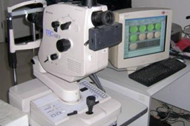 Corneal pachymetry is a measurement of the thickness of the cornea. It can be performed using contact methods, such as ultrasound, or non-contact methods such as Optical Coherence Tomography (OCT). Corneal pachymetry is particularly essential prior to a LASIK procedure.
Electrooculography (EOG/E.O.G.) is a technique for measuring the resting potential of the retina. The resulting signal is called the electrooculogram. The main applications are in ophthalmologic diagnosis and in recording eye movements. Recording of the average amplitude of the resting potential arising between the cornea and the retina in light and dark adaptation as the eyes move a standard distance to the right and the left. The increase in potential with light adaptation is used to evaluate the condition of the retinal pigment epithelium.
Computerized Corneal topography, also known as photokeratoscopy or videokeratography, is a non-invasive medical imaging technique for mapping the surface curvature of the cornea, the outer structure of the eye. This method could be employed for diagnostics such eye conditions as keratoconus, also in planning refractive surgery such as LASIK and evaluation of its results or in assessing the fit of contact lenses. Since the cornea is normally responsible for about 70% of the eye's refractive power, its topography is of critical importance in determining the quality of vision.
Tests
Following to the official medical regulations all patients before surgery should undergo a preoperative preparation, including blood tests (AIDS, RW, Hepatitis B and C), urinalysis, electrocardiography(ECG) and a consultation of anesthesiologist. The preoperative tests and analysis are used to detect previously unknown conditions that may influence surgical outcomes. Urinalysis can reveal diseases that have gone unnoticed because they do not produce striking signs or symptoms. Examples include diabetes mellitus, various forms of glomerulonephritis, and chronic urinary tract infections. The electrocardiogram (ECG or EKG) is a diagnostic tool that is routinely used to assess the electrical and muscular functions of the heart. All tests and analysis are available at the “OkoMed” eye clinic and take a minimum of time.
Contact Lenses correction
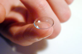 Contact lenses are used to correct the same eye conditions that eyeglasses do: myopia (near or short-sightedness), hyperopia (far or long-sightedness), astigmatism (blurred vision due to the shape of the cornea) and presbyopia (inability to see close up).
There are two general types of contact lenses: hard and soft. The hard lenses most commonly used today are rigid, gas-permeable lenses (RGP for short). Soft lenses are the choice of most contact lens wearers. These lenses are comfortable and available in many versions, depending on how do you want to wear them.
Contacts could be:
- Daily-wear lenses;
- Extended-wear lenses;
- Disposable-wear lenses;
- Cosmetic or decorative contact lenses;
- Toric soft contact lenses can correct astigmatism;
- Bifocal or multifocal contact lenses are available in both soft and RPG varieties.
Contact lenses if are not properly cleaned and disinfected increase the risk of eye infection. Any contact lens that is removed from the eye needs to be cleaned and disinfected before it is reinserted. Our eye care professional will discuss the best type of cleaning system for particular patient, depending on the type of lens patient uses.
Eyeglasses fitting
After very careful vision examination, only ophthalmologist prescribes eyeglasses for your vision correction. Skilled experts of the clinic “OKOMED” fit eyeglasses of any complexity and purpose (spectacles for reading, for computer, for driving, for permanent wearing and others). In addition, any patient could choose a modern eyeglass frame and order making spectacles.
We use and offer to our patients eyeglass lenses of the world famous manufacturers who have quite number of protective coatings, anti-reflective including (spectacles for computer).

