Conventional Eye Microsurgery
Cataract surgery in our clinic is performed by the method of micro incision cataract surgery (MICS) with ultrasonic phacoemulsification and implantation of intraocular lenses (IOLs).
The cataract is one of the most common eye disorders among people of old age. This eye disease connected with opasification of the natural human crystalline lens. Cataract leads to partial or complete dimness of the crystalline lens, thus, only part of the light is able to reach the retina. This means that the affected person has poor vision and the images are unclear and blurred. There are several types of cataract such as: congenital cataract, traumatic cataract, complicated cataract, radiation cataract, and the disorder caused by general diseases. More often, the cataract is age-related and develops in people over 50.
With years, the disorder develops, the area of dimness grows and vision deteriorates further. Failure to undergo cataract treatment in time may lead to complete blindness. Up-to-day technology allows to treat this disorder at any stage and patient could not suffer from poor vision, waiting when cataract became “mature.”
In “OkoMed” eye clinic we use the latest model of phacoemulsificator the INFINITI® Vision System (Alcon, USA) and ultra-thin flexible, the best available today, intraocular lenses of the "PREMIUM class” from the leading manufacturers of the UK, USA and Germany such as Aspheric Ultima, Rayner Superflex, Aqua Free and multifocal M-Flex Rayner (UK), Aspheric Foldable Bioline Yellow and multifocal AT LISA (Germany), multifocal AcrySof IQ Natural, AcrySof IQ toric and aspheric, AcrySof IQ ReStor, Alcon (USA).
The INFINITI® Vision System is the first system with three levels of impact energy to the lens: AquaLase ®, NeoSoniX ® and ultrasound modulation. This technology combines an extraordinary fluidics with two unique energy delivery systems. The system's OZIL® Torsional Handpiece delivers side-to-side, oscillating ultrasonic movement, increasing control of the surgeon during procedure.
Multifocal IOL restored distance and near vision without glasses, toric lenses - correcting astigmatism. Phacoemulsification refers to modern cataract surgery in which the eye's internal lens is emulsified with an ultrasonic handpiece and aspirated from the eye. Aspirated fluids are replaced with irrigation of balanced salt solution, thus maintaining the anterior chamber, as well as cooling the handpiece.
Non penetrating deep sclerectomy
Non penetrating deep sclerectomy (NPDS) also called non penetrating trabeculectomy, is a filtering surgery where the internal wall of Schlemm's canal is excised, allowing subconjunctival filtration without actually entering the anterior chamber. NPDS appears to be an effective and safe filtering procedure for lowering IOP and could be an alternative to trabeculectomy in open angle glaucoma with the advantage of having fewer complications.
Deep sclerectomy with allodrainage
This surgery is indicated for most of glaucoma except angle closure and neovascular cases. The procedure consists in opening the conjunctiva and Tenon’s capsule and creating a 5 × 5 mm limbus-based superficial scleral flap. A deeper scleral flap measuring about 4 × 4 mm is dissected and the roof of Schlemm’s canal is removed. A space maintainer is inserted and the flap and conjunctiva are closed.
Vitrectomy surgery with full range of surgical manipulations:
- Endolasercoagulation;
- Separation of commissure of the vitreous body;
- Blocking of retinal breaks and holes;
- Introduction of perfluorocarbon, silicon.
Since 2012 our clinic has successfully performed vitreoretinal surgery of the disorders of the posterior segment of the eye.
Vitrectomy (removal of the vitreous body) by access through a microscopic incision of the eye at the flat part of the ciliary body using the superfine tools allows to treat successfully vitreous opacifications, haemophthalmos, retinal detachment, combination of the retinal detachment with the pathology of vitreous and lens.
Vitrectomy with removal of the newly formed membranes and commissure near the retina is performing in the case of epiretinal membranes (inner limiting membrane peeling), vitreo macular traction syndrome, macular retinal tears, diabetic macular edema, edematous form of age-related macular dystrophy.
Special technologies are used during vitrectomy surgery as a supplement to this technique: endolasercoagulation, blocking retinal breaks and holes, separation of commissure of the vitreous body , the introduction of gas, perfluorocarbon and silicone.
New special surgical microscope with additional equipment produced by"DORC" company (Germany) allows to remove the vitreous body and to conduct complex manipulations near the retina to remove membranes, commissures and fibrosis using endolasercoagulation.
Macular dystrophy of the retina surgery
Macular dystrophy is a genetic eye disorder that can cause deterioration of central vision and eyesight loss. This disorder affects the retina, specifically cells in a small area near the center of the retina called the macula. The macula is responsible for sharp central vision, which is needed for detailed tasks such as reading, driving, and recognizing faces.
Retinal detachment surgery
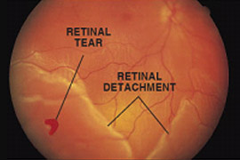
 Symptoms of a detached retina include sudden visual changes such as dark spots, flashing lights and shadows in the vision. Retinal detachment surgery involves reattaching the retina to the back of the eye and sealing any breaks or holes in the retina. If a detached retina is not treated it can lead to blindness. Is it possible to restore vision in such cases mostly depends on the extent and location of the retinal detachment, mainly this type of surgery is intended to save the rest vision than to improve it.
Strabismus (Squint or Cross-Eyes) surgery
Strabismus may be concomitant or incomitant (paralytic). It could be convergent strabismus (esotropia) in which one eye turns inward, divergent strabismus (exotropia) in which one eye turns outward, vertical strabismus in which one eye turns upward.
When the eye turn occurs all of the time, it is called constant strabismus. When the eye turn occurs only from time to time, it is alternating strabismus.
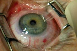
Strabismus surgery strengthens or weakens eye muscles, which changes the alignment of the eyes relative to each other. Eye muscle surgeries typically correct strabismus and include the following:
Loosening / weakening procedures:
- Recession involves moving the insertion of a muscle posteriorly towards its origin;
- Myectomy;
- Myotomy;
- Tenectomy;
- Tenotomy.
Tightening / strengthening procedures:
- Resection involves detaching one of the eye muscles, removing a portion of the muscle from the distal end of the muscle and reattaching the muscle to the eye;
- Tucking;
- Advancement is the movement of an eye muscle from its original place of attachment on the eyeball to a more forward position;
- Transposition / repositioning procedures;
- Adjustable suture surgery is a method of reattaching an extraocular muscle by means of a stitch that can be shortened or lengthened within the first post-operative day, to obtain better ocular alignment.
High degree myopia surgery To correct high degree myopia (near or shortsightedness) we are using eye surgery method of implantation of phakic intraocular lens (PIOL) - so called “glasses inside eyeball”, also called implantable contact lenses (ICLs), which have been shown to be a safe and effective means of correcting moderate to severe myopia. Intraocular lens is called “phakic” because the eye's natural lens is left untouched. ICL's or “phakic” implants, which retain the natural lens of the eye.
Phakic intraocular lenses are indicated for patients with high refractive errors when the usual laser options for surgical correction such as (LASIK and PRK) are contraindicated. Phakic IOLs are designed to correct high myopia ranging from -5 to -20 D if the patient has enough anterior chamber depth (ACD) of at least 3 mm.
Three types of Phakic IOLs are currently available:
- Angle-Supported;
- Iris-Fixated;
- Sulcus-Supported: Implantable Collamer Lens (Visian ICL) is a type of sulcus supported phakic intraocular lens used to treat myopia between -3D to -20D and for people between 21-45 years of age.
Phakic intraocular lenses, are a new option for people seeking more permanent correction of common vision errors such as myopia (nearsightedness).
These implants, which resemble contact lenses, are placed in either of two locations in the eye. The first one is between the cornea and the iris and the second one is just behind the iris. Unlike traditional contact lenses, you cannot feel a phakic IOL in your eye. Also, phakic IOLs require no maintenance.
The treatments can differ slightly and be as follows:
- A Fixed Focus lens implant: This type of lens is recommended if the patient is over 40 and doesn't using reading glasses, or if the patient has cataracts;
- A Fixed Focus lens implant is also used when patients suffer from a specific type of refractive disorder, including high myopia;
- A Monovision lens implant: This lens implant enables the patient to see both near and far away without the need of any additional correction. It addresses one eye for near vision and the other for distance vision, giving the patient a full range.
Refractive Lens Replacement Surgery (RLR), in which the eye's natural lens is replaced.
In certain cases we could use the method of exchange (replacement) of the natural lens for IOL of less optical power.
High degree hyperopia surgery
By the method of exchange of the natural lens for artificial IOL of higher optical power we successfully treat hyperopia (far-sightedness) of high degree and get 100% visual acuity without correction.
Multi-Focal lens implants: Presbyopic Lens Exchange (PRELEX) implants are part of a technique used to treat presbyopic and hyperopic patients. This technique is not recommended for patients with small pupils or those who do a lot of night driving due to the possibility of halo effect.
Progressive myopia surgery
To stop the process of progression of the axial length growth of an eyeball in myopia we offer scleroplasty surgery.
Collagenoplasty, these are collagen injections to stop the progression of myopia. Collagen stimulates special scleral cells and they produce a new scleral tissue that prevents scleral stretching and finally stop growth of the eye ball.
Eyelids, Conjunctiva & Lachrymal surgery
Our clinic provides full range of ophthalmologic treatment of eyelids, conjunctiva and lachrymal organs disorders:
- Removal of pterygium;
- Chalyazion surgery;
- Removing tumors of eyelid or conjunctiva;
- Setting obturators of lachrymal pathway;
- Surgical enlargement of lachrymal points;
- Probing of lachrymo-nasal canal;
- Removing molluscum contagiosum;
- Dissection of retention cysts of eyelids and conjunctiva;
- Removal of stye (an external stye or sty, also hordeolum);
- Removing "millet seeds";
- Eyelashes epilation;
- Plastic on the lower eyelid by the method of Shotter in case of it’s age spastic blepharelosis.
Pterygium (Surfer's Eye) most often refers to a benign growth of the conjunctiva. A pterygium often grows from the nasal side of the sclera. It is usually present in the palpebral fissure and can be caused by ultraviolet-light exposure (e.g., sunlight), low humidity, and dust. The predominance of pterygium on the nasal side is possibly a result of the sun's rays passing laterally through the cornea, where it undergoes refraction and becomes focused on the limbic area. On the contra lateral (medial) side, however, the shadow of the nose medially reduces the intensity of sunlight focused on the lateral/temporal limbus. Pterygium can be removed surgically for cosmetic reasons, as well as for functional abnormalities of vision or discomfort.


